The horse digestive system is an impressively complex labyrinth—about 100 feet long stem to stern—that plays no small part in your horse’s overall health. And while you may know your colic from your colitis, how well do you really know the ins, outs, and in-betweens of your horse’s digestive system? Read more and get to know your horse’s gut.
Mouth
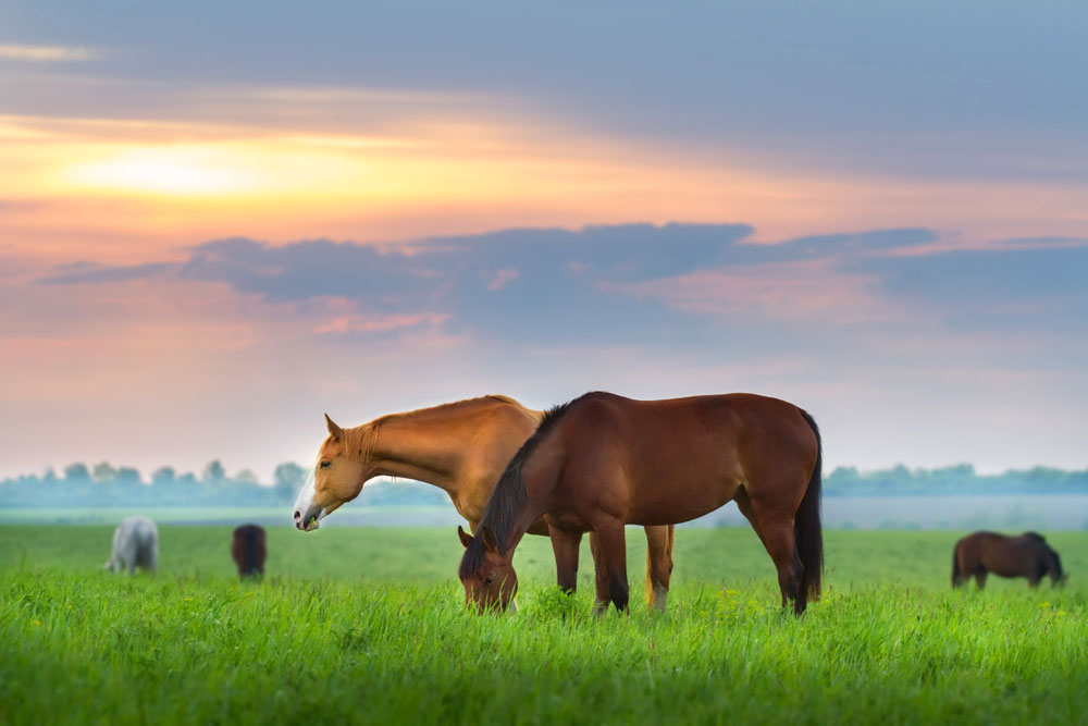
Your horse also has large salivary glands that produce up to 10 gallons of saliva per day. This helps moisten the food before swallowing. Saliva also contains an enzyme called amylase, which begins the digestive process even before food is swallowed.
Esophagus
This long, muscular tube is roughly 4 to 5 feet long in an adult horse and takes feed from swallow to stomach. The esophagus pushes food down in a wave-like motion called peristalsis. If you watch closely, you can sometimes see a lump of feed work its way down a horse’s neck.
Occasionally, a chunk of feed will get stuck in the esophagus—this is referred to as “choke,” although that’s a bit of misnomer, since a horse with an esophageal obstruction can still breathe.
Choke can be dangerous; the esophagus is relatively thin-walled and can be damaged and scarred, or even torn. A horse with choke is also at risk for developing aspiration pneumonia due to accidental inhalation of food particles or water.
A horse with choke will appear distressed and may play in water but not drink, or repeatedly stretch out his neck and attempt to swallow. Medical intervention is typically required to relieve choke. Your veterinarian will sedate the horse and create a siphon with a nasogastric tube to lubricate and break up the blockage through direct irrigation and rehydration with water.
Stomach
The esophagus enters the stomach at an extremely acute angle. This unique anatomical feature of the horse prevents ingesta from easily coming back up, therefore preventing horses from being able to vomit.
Horses’ stomachs are relatively small—holding only 2 to 4 gallons—and they empty very quickly. If horses aren’t allowed to eat frequent, small meals, the rapid emptying of the stomach leaves the lining exposed to gastric acid, and ulcers can form.
Small Intestine
After a quick exit from the stomach, food enters the small intestine. About 70 feet in length with a total capacity of about 12 gallons, the small intestine has three parts, as in humans: the duodenum, jejunum, and the ileum.
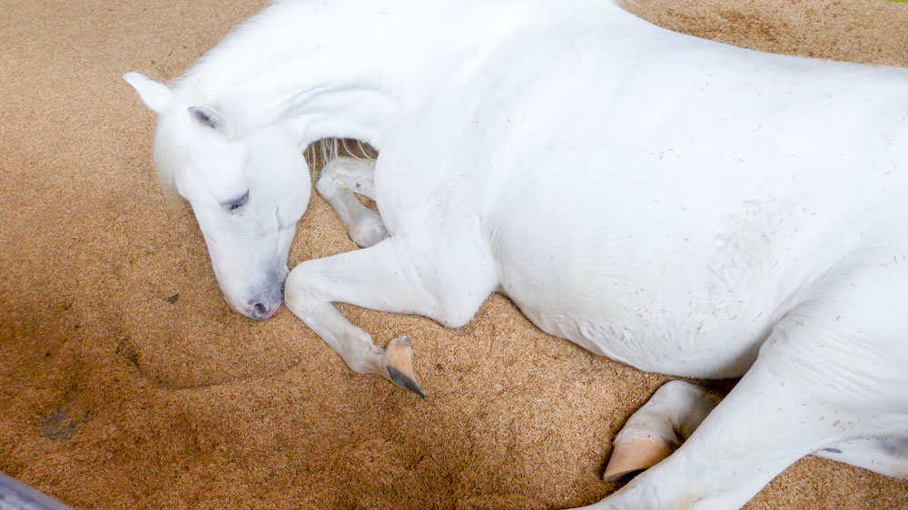
Throughout the small intestine, serious digestion takes place as numerous types of digestive enzymes are secreted. The pancreas also contributes various enzymes, and bile from the liver is added in as well.
With abundant mixing, carbohydrates, proteins, and fats are broken into their smaller subunits and absorbed through the walls of the small intestine.
Large Intestine
Up to this point, fiber from roughage such as grass and hay has not been broken down. The primary functions of the horse’s large intestine (made up of the cecum, large colon and small colon) are to house microbes that break down cellulose via fermentation and to absorb water and vitamins.
CECUM:
The first part of the large intestine is the cecum. Food enters the cecum, a large, comma-shaped organ on the right side of the horse’s abdomen, after exiting the ileum. It holds roughly 8 to 10 gallons and it is here where fermentation truly starts.
Horses are sometimes referred to as “hind gut fermenters” for this reason. This is in contrast to cattle, which begin fermentation much earlier in the digestive process, in their rumen (four-chambered stomach).
The cecum in the horse is oddly shaped; it is a blind-ended sac with food entering and exiting near the top. Occasionally, if a horse is dehydrated, an impaction can occur in the far end of the cecum, causing colic. This is called a cecal impaction.
Feed can remain in the cecum for several hours, during which time microbes ferment the abundant cellulose.
LARGE COLON:
After this, food starts its long journey through the large colon. This tube lives up to its name; at about 12 feet long and 10 inches in diameter, this organ doubles over itself to form two U-shapes along the perimeter of the horse’s abdomen.
The cecum and large colon are common locations for parasites in horses. Small strongyles (cyathastomes) encyst within the walls of these organs. Large numbers of these cysts can cause extensive damage when the parasites emerge, causing reduced nutrient absorption and subsequent weight loss and diarrhea.
Large strongyles also migrate to the large intestine. Strongylus vulgaris is the nastiest of these parasites, because it not only damages the intestinal wall, but also on further migration, moves up the cranial mesenteric artery, potentially blocking the blood supply to parts of the colon, resulting in colic.
From the cecum, food enters the right ventral colon and moves toward the front of the horse. Within the colon, there are several narrow turning points called flexures. As food moves from right to left ventral colon, it passes through the sternal flexure, named because it’s a turn near the sternum.
Moving along the floor of the abdomen now on the left side, food then encounters the pelvic flexure (back near the pelvis) and then up to the section of colon sitting on top of itself. The pelvic flexure can be felt during rectal palpation and is another common location for impactions due to its hairpin turn.
After moving through the pelvic flexure, food enters the left dorsal colon and heads toward the front of the horse again. After this, food passes through the last narrow turn called the diaphragmatic flexure, then into the right dorsal colon. This is where an overdose of NSAIDs such as phenylbutazone can cause right dorsal colitis, an irritation of the lining of the colon, resulting in diarrhea and intermittent colic.
SMALL COLON:
Leaving the right dorsal colon, food enters the relatively short transverse colon and into the small colon, also known as the descending colon. This is the last stage of the digestive process.
All nutrients have been removed at this point, and the primary function of the small colon is to absorb any remaining moisture. The distinctive balls of horse manure are formed here before moving through the rectum and out of the animal.
The entire digestive process in the horse can take anywhere from 36 to 72 hours, depending on amount and content of the feed.
This article on the horse digestive system originally appeared in the May 2018 issue of Horse Illustrated magazine. Click here to subscribe!
Further reading:
Nutrition: The Key to Unlocking Your Horse’s Health
Hay Quality & Nutrition: Evaluating Your Horse’s Nutritional Needs

