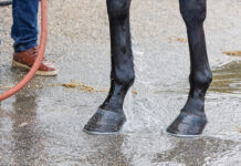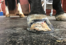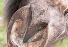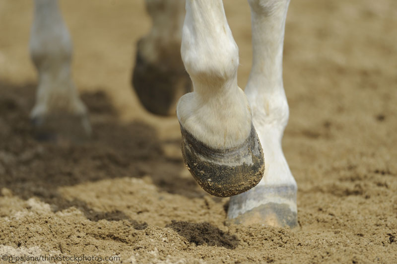
But what happens when the next word you hear is “navicular?” Is it a dire prediction of permanent lameness, or simply a realistic location of temporary pain?
A lameness examination shouldn’t trigger a panic attack. Navigating a potential diagnosis of navicular-area lameness is no different from driving successfully through a complicated street intersection: Look for signs, steer clear of distractions, and keep both hands on the wheel.
The Structures
Start by thinking of the navicular bone in your horse’s foot like a statue in the middle of that intersection. This tiny bone sits in a critical spot near the middle of the foot. (Did you know there’s a navicular bone in your own foot, as well?)
The bone is held in place with multiple ligaments. Behind it sits a bursa (sac) that cushions it from the tendon behind it. The foot’s blood supply and nerves weave through it all like busy bike messengers through that intersection; the sole, frog and digital cushion are underneath it. This complicated design works; everything functions cooperatively in a sound horse.
If your horse is lame, be patient until you have a definitive diagnosis and the all-important prognosis or recovery forecast. That statue in the intersection may not be damaged after all, but instead, structures around it are stressed or injured. Most damage can heal.
What happens next? Expect tests, radiographs, and possibly more advanced imaging before your veterinarian’s final report. Your horse will be resting. Veterinarians often start horses on pain-relieving anti-inflammatory medication; later plans might call for a more advanced treatment, like bisphosphonate medication. Joint injection is a possibility, as are new shoes (or no shoes at all).
Horses with similar signs of hoof lameness often have different injuries and require very different treatments. The age and use of the horse can also dictate what treatment is prescribed. Some owners want their horses back in the arena as soon as possible, while others are happy to give a horse time off or accept a future change in use.
Be prepared for questions your veterinarian may ask about your horse’s training schedule, any changes made recently, and details of past lamenesses.
Navicular Syndrome
Horses go lame every day, for many reasons. But one of the most common reasons is because of lameness in the region of the foot where the navicular zone is located. In fact, veterinarians estimate that about 30 percent of lameness is what we’ll call “navicular-ish.”
Veterinarians have studied navicular-type lameness for at least 250 years. For an equal time, farriers tried to make those lame horses comfortable. Both professions have learned a lot. Medications have been developed to ease navicular lameness. Dozens of products have been marketed.
But horses still go lame, and veterinarians still say, “Well, it could be his navicular. We need to look further. Don’t panic.”
But you do, anyway.
Not too long ago, horse owners feared the serious foot lameness called navicular disease, and many still do. Horses have been retired or even euthanized when therapy and medication failed to restore soundness.
Veterinarians were confounded to learn that some sound horses had damage or degeneration in the bone, but no sign of lameness. Some lame horses had clean radiographs.
Thanks to the availability of radiographs in the field beginning in the 1970s, veterinarians were able to compare the images of lame horses. Horses with similar lameness symptoms might not have similar radiographs.
Eventually, navicular disease began to be thought of as a “syndrome,” or group of related symptoms. Case analyses showed that Quarter Horses and European warmblood breeds were commonly affected.
Horse Navicular Disease: Current Diagnostics
Technically, “navicular disease” is a specific chronic degeneration of the navicular bone itself, and it still exists, but veterinarians are very careful before they diagnose it.
With the advent of magnetic resonance imaging (MRI) for horses, the maze of structures within the foot was finally visible from every angle—but an MRI might show multiple problems in the same foot.
Today, some veterinarians simply refer to “palmar foot pain” until or unless a definite diagnosis can be made. They are describing lameness symptoms in the back of the foot; palmar foot pain is not a diagnosis.
A diagnosis identifies an illness or injury; the exact cause of palmar foot pain can be difficult and expensive to ascertain. A series of nerve block diagnostic tests uses temporary local anesthetic injections to narrow down the source of pain.
Sometimes—but not always—the ultimate diagnosis will still be the true degenerative “navicular disease.” It’s a frustrating diagnosis, since some horses respond initially to treatment only to have the lameness return. As a last resort, some horses with navicular pain undergo nerve-cutting surgery to desensitize the navicular zone and alleviate pain.
“In navicular disease, you treat the pain, not the lameness, because the damage is irreversible,” says Jean-Marie Denoix, DVM, Ph.D., Ass. LA-ECVDI, DECVSMR (Equine), DACVSMR (Equine), of the National Veterinary College in France.
Pinpointing what causes navicular-type foot pain is still the challenge. Quite often, “wait and see” or “if this doesn’t work, we’ll…” are heard just as often as they were years ago.
Some horses respond positively to simple changes in workload, arena surface or even warmup routines. Some can go back to work on light medication. Other horses are less responsive.
Name that Lameness
Palmar foot pain covers the big picture of discomfort in the navicular zone, sole and heels. Digging deeper, we can see how complex—and co-existing—the different lameness problems can be.
A study led by Sue Dyson, MA, VetMB, Ph.D., DEO, FRCVS, clinician at England’s Animal Health Trust equine orthopedics referral center, examined the MRI records of 263 horses with foot pain. Only six showed abnormalities in the navicular bone alone; 29 had injury to both the bone and the tendon. Another 60 had co-existing injury in the bone, navicular ligaments and/or tendon.
They documented 46 horses with injuries to the collateral ligament of the coffin joint in combination with lesions of the tendon, navicular ligaments or navicular bone. Still another 25 horses had damage in five or more structures within the foot.
Horses sometimes show signs of foot lameness when they have been changed to different shoes, or a different farrier. A recent change in owner, rider, exercise intensity, or tack is worth pondering during early stages.
Something as simple as a bruised foot or sore heels can cause temporary palmar foot pain. Injuries like collateral ligament desmitis, pedal osteitis, coffin joint arthritis, or infected puncture wounds also cause pain in this part of the foot.
MRI has made it possible to diagnose damage to the deep digital flexor tendon; veterinary radiologists will look for tiny tendon tears in the foot to see if they (or more general tendonitis) are possibly at the root of pain. The navicular bursa, the small sac protecting the navicular bone from the tendon, can also be damaged.
Horse Navicular Disease: Corrective Farriery
The first step for many horses with palmar foot pain is referral for therapeutic farriery. Some horses with palmar foot pain suffer from overuse, long show seasons, riding on hard ground, conformational challenges and poor or infrequent trimming and shoeing.
Their feet may have weak walls or cracks; the foot may be contracted and the heels underrun. Restoring good hoof balance will be a top priority and may work wonders.
Make sure your farrier plans to come on a schedule to re-trim or re-shoe your horse through his rehab period. The farrier may need to come every four weeks instead of every six to eight weeks.
Radiographic evaluation of farriery is a new possibility; for this convenience, your horse may need to be shod at a vet clinic, or both the farrier and the veterinarian must visit your barn at the same time.
The veterinarian will radiograph the horse before and after shoeing. The support or relief of different areas of the foot can be evaluated, along with the improved balance of the foot.
Does your farrier know your horse well? Farriers know what worked for individual horses in the past; they will watch how horses move and design or modify shoes that minimize effort or fully support the foot.
Your horse will also let a farrier know when he doesn’t like a shoe. After shoeing, the horse may change the way he stands or shorten his stride; your farrier can interpret signs that the horse wants more stability than a rocker shoe offers, or less heel elevation than prescribed wedges.
Watch your horse carefully after he has been shod. Sometimes putty-like hoof packing is uncomfortable for one horse, while another horse shows a positive response; this is all vital information for your team to know.
When your horse is lame with navicular-ish pain, you’re sharing a common experience with horse owners around the world. Remember that most horses respond to treatment and rest.
This article about horse navicular disease originally appeared in the July 2018 issue of Horse Illustrated magazine. Click here to subscribe!

